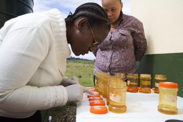
BRIGHT Academy research

Female genital schistosomiasis is currently not diagnosed by health systems in any country. This means that unknowing doctors and nurses are mistakenly treating and advising female patients as though they have a sexually transmitted disease. Millions of women are untreated for female genital schistosomiasis. Millions of young girls grow up into adulthood with a disease that could be prevented.
Diagnosing Female Genital Schistosomiasis (FGS)
Visual examination by health professionals – we started this with the World Health Organisation
Increased awareness amongst health professionals and patients is a prerogative for correct management. Together with the WHO and a number of gynaecologists we have therefore made
- an atlas for genital schistosomiasis that is freely available online and
- a pocket atlas and
- a freely downloadable bedside poster
Diagnostic support – we are working on this, please be impatient!
It is highly likely that FGS could be diagnosed by automatic analysis of the colour of the lesion (Holmen 2015-16). Although some lesions are different in Madagascar and mainland Southern Africa the colour and the abnormal blood vessels are the same, it seems. With our partners in technology and cervix cancer prevention programmes we are in the process of creating a joint diagnostic tool. Why waste the opportunity to check for both diseases when a patient has made the effort to come?
Female Genital Schistosomiasis (FGS) as a risk factor for HIV
Women who do not have access to safe water, such as the inhabitants in rural Africa may have genital lesions caused by the parasite Schistosoma haematobium (Bilharzia). Studies in Zimbabwe and Tanzania have shown 3 to 4-fold higher prevalences of HIV in women with FGS. There is increasing evidence that FGS is a co-factor in HIV-transmission, similar to the sexually transmitted diseases. We hypothesize that standard treatment can prevent HIV-transmission in young women. Substantiation of the link between HIV and schistosomiasis and the biological mechanisms behind it would provide a novel intervention point against HIV transmission through implementation of the recommended mass treatment. Importantly, information should be given to the patient about her risks.
Pathogenesis and clinical presentations of FGS
Genital lesions caused by the parasite Schistosoma haematobium (Bilharzia) are commonly found in inhabitants in rural Africa where the people do not have access to clean water for domestic use and for play. Through our work in Zimbabwe we unexpectedly found that in adult women, genital lesions were calcified and standard treatment was ineffective. We have limited knowledge about the clinical and histological courses of genital S. haematobium infection. What does an early and late lesions look like? We will also explore the impacts of S. haematobium and S. mansoni on fertility and cancer. BRIGHT Academy addresses the clinical gaps and the unresolved research questions.
Treatment of FGS
Praziquantel is a potent anti-helminthic drug, which kills the worms but has limited effect on pathology that was established by egg deposition a long time ago. Therefore, once schistosomiasis lesions (visualised as so-called sandy patches) are established, the lesions and the surrounding inflammation, persist despite treatment. In fact, it has been shown that treatment has limited effect on the disease in adult women. Interestingly, however, a retrospective study showed that women who had received anti-schistosomal treatment in childhood had significantly fewer lesions and less contact bleeding (Hegertun et al 2013). In order to prevent the morbidity caused by repeated re-infection, praziquantel must likely be administered several times in childhood. The pivotal work in this programme is to explore if treatment has an effect in young women. The optimal age for treatment with praziquantel is unknown.
